Medial Collateral Ligament Injury Physiotherapy Treatment
Medial collateral ligament injury
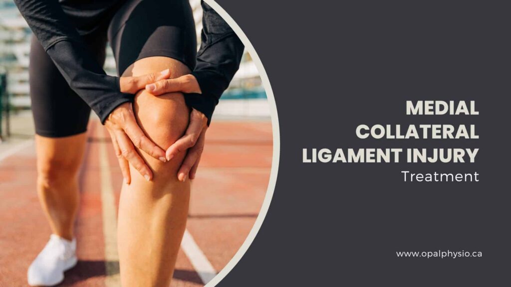
The Medial collateral ligament (MCL) is a crucial component of the medial knee, providing stability and preventing the joint from moving from side to side. MCL injuries commonly occur due to direct impact or force that pushes the knee inward, leading to stress or tearing of the ligament.
An MCL injury can range from a mild sprain to a complete rupture, significantly impairing mobility and causing considerable discomfort in the knee.
Medial collateral ligament injury is the most common knee injury seen in athletes. Understanding the causes, symptoms, risk factors, and appropriate treatment options, including physiotherapy, is crucial for managing MCL injuries effectively.
MCL injury physiotherapy treatment in Langley
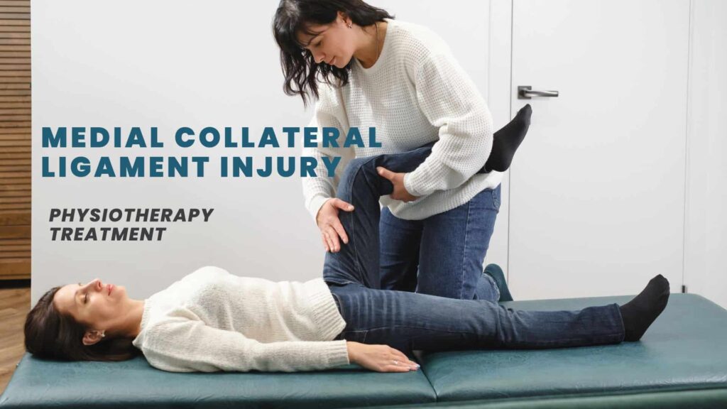
Physiotherapy treatment for MCL injury can help reduce pain and swelling and improve knee stability. Treatment may include exercises to strengthen the muscles around the knee and improve the range of motion.

What is a medial collateral ligament?
The medial collateral ligament (MCL) is a tissue band that runs down the inner side of the knee. It connects the femur( thigh bone ) to the tibia ( shinbone )and helps to stabilize the knee joint. The medial collateral ligament is one of the four major ligaments stabilizing the knee joint. The MCL is responsible for preventing the knee from collapsing inwards (valgus force) and for providing stability to the knee when the leg is flexed.
Causes of medial collateral ligament injury
MCL injuries are typically caused by
In addition to acute injuries, the medial collateral ligament can be damaged over time through repeated stress and pressure, such as lifting heavy objects.
Symptoms of MCL injury
The symptoms of an MCL injury can vary based on the severity of the tear. Most people who tear their MCL feel a “pop” in their knee when the injury happens. The symptoms can include
Risk factors for MCL injury
Certain factors can increase the risk of an MCL injury. These include:
Medial collateral ligament injury treatment options
Most MCL injuries don’t require surgery, and a non-surgical approach is often preferred initially. It can be treated at home with rest, ice, and anti-inflammatory medicine. In some cases, crutches and a knee brace may be recommended to reduce further strain. Physical therapy is also a common part of the treatment plan to help with strength and flexibility after the injury.
In severe cases, such as a grade 3 tear where the ligament is completely torn, surgery may be required.

Physiotherapy treatment for medial collateral ligament injury
Physiotherapy plays a crucial role in the recovery from a medial collateral ligament injury. The goal is to rebuild the strength of the knee muscles and regain motion. This can involve exercises such as stationary cycling and specific strength and flexibility exercises, including
Recovery timeline
MCL injuries vary in severity, but most individuals can return to normal activities within a few weeks or months with proper care. Severe cases may require a more extended recovery period.
It should be noted that in the initial phase of recovery, the knee should be protected with a short-hinged brace for 3 to 6 weeks, depending on the severity of the injury. Crutches and restricted weight bearing may also be needed.
Remember, it’s essential to consult with a healthcare professional or physiotherapist to determine the best treatment plan for your specific injury.
Opal Physiotherapy Clinic provides physiotherapy treatment for medial collateral ligament injuries in Langley. Our team of physiotherapists will develop a personalized treatment plan for you based on the severity of your injury.
Our experienced and qualified physiotherapists are dedicated to helping our patients recover from their injuries. We will work with you to help you regain full function of your knee and get you back to your normal activities as soon as possible.
If you have a medial collateral ligament injury, seeing our physiotherapist as soon as possible is important. Early treatment can help to reduce the risk of further injury and to speed up the healing process.
